In chronic wound management, “perfusion” is often limited to pulse or vessel examination alone. Clinically, perfusion is blood flow reaching the wound bed and surrounding tissue, and with it the delivery of oxygen and nutrients. Granulation tissue formation, epithelialisation, and capacity to manage infection depend on adequacy at this microcirculatory level. If perfusion is inadequate, healing does not progress at the expected rate no matter how thoroughly the wound surface is cleaned. This article summarises the clinical methods used to assess the blood supply to the wound and surrounding tissue — venous versus arterial distinction, ankle–brachial pressure index (ABPI), and transcutaneous oxygen pressure (TcPO2).
Perfusion: nutrition of wound tissue and surrounding skin
Wound healing is not limited to cells in the wound bed. The wound margin, surrounding skin, and underlying soft tissue also depend on the same perfusion network. If arterial inflow is inadequate, tissue oxygen falls; if venous outflow is impaired, oedema and metabolic waste accumulation increase. In both cases, granulation quality weakens even when the wound bed looks “hungry.”
At the microcirculatory level, perfusion means capillary refill, oxygen diffusion, and clearance of metabolites such as lactate. Clinically we assess this indirectly through skin colour, temperature, oedema, pain, margin viability, and healing rate. Pulse palpation is valuable for screening; yet in diabetic foot and calcified vessel profiles, distal tissue nutrition may remain inadequate even when the pulse feels “normal.”
In chronic venous insufficiency the problem is often venous hypertension and rising capillary pressure: tissue nutrition is impaired by venous stasis, and oedema leads to protein-rich exudate. In arterial ischaemia the problem is on the inflow side: oxygen and nutrients do not reach the wound base adequately. Both mechanisms can coexist in the same leg; perfusion assessment then has to address both mechanisms.
Venous and arterial profiles: impaired blood supply behaves differently
Venous ulcers are common on the lower extremity in the malleolar and gaiter region. Hyperpigmentation, lipodermatosclerosis, varicose networks, and oedema show that surrounding tissue remains under venous stasis. The wound margin is irregular; exudate may be moderate to high. Clinical stage is recorded with CEAP classification; C5–C6 is open venous ulcer, C4 is pre-ulcer skin change.
In the arterial profile the wound is usually distal — on the toes or pressure points. Skin is cool, pale, or cyanotic; rest pain often accompanies. The wound margin may look “punched out”; necrotic tissue is prominent. Peripheral artery disease (PAD) and critical limb ischaemia (CLI) represent the arterial face of perfusion failure in this picture.
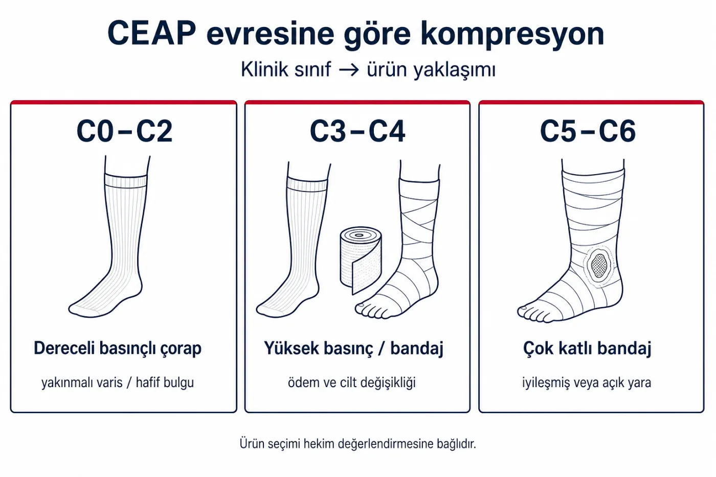
In both profiles the question is the same: is there enough oxygen and nutrient at the wound bed and margin for healing? In the venous patient the answer usually relates to oedema control and lowering venous pressure; in the arterial patient, raising inflow pressure and distal perfusion comes to the fore.
ABPI: arterial inflow screening
Ankle–brachial pressure index (ABPI) is the ratio of ankle systolic pressure to arm pressure. It is measured with Doppler; in practice the 0.9–1.3 range is accepted as normal in most guidelines. 0.5–0.9 suggests moderate disease; below 0.5 suggests critical ischaemia. Values above 1.3 may be misleading because of calcified vessels.
ABPI is the most common tool for screening arterial inflow adequacy around the wound. When compression is planned in a venous ulcer profile, arterial adequacy is assessed first; external pressure can further reduce inadequate arterial inflow. The compression pressure (mmHg) article addresses ABPI and contraindication summary further in the venous ulcer context.
When ABPI alone is not enough
- Diabetes and chronic kidney disease — medial calcification can elevate the index.
- Distal wound (toes) — ankle pressure may not reflect toe perfusion.
- Severe oedema — cuff placement is difficult; measurement is repeated.
In these profiles toe–brachial index (TBI) or TcPO2 provides complementary information. Revascularisation decisions are not reduced to a single ABPI value; clinical picture, imaging, and tissue-level measurements are considered together.
TcPO2: tissue-level oxygenation
Transcutaneous oxygen pressure (TcPO2) is partial oxygen pressure measured at the skin surface with a heated electrode. Unlike macroscopic pulse and ABPI, it reflects tissue-level oxygenation and is used to assess nutritional adequacy of the wound bed and surrounding skin.
Threshold values are set in line with institutional protocol and guidelines. In general, TcPO2 below 30 mmHg suggests inadequate tissue oxygenation for healing; above 40 mmHg is associated with more favourable perfusion. Standing and supine measurements may differ; position should be noted in the report.
TcPO2 complements ABPI in diabetic foot and mixed-aetiology wounds. Serial measurement before and after revascularisation gives vascular surgery and the wound team a common reference. Healing expectations for the wound bed cannot be framed realistically until distal perfusion is clear.
Clinical flow in perfusion assessment
Perfusion assessment during wound examination follows this sequence:
- Aetiology — venous, arterial, neuropathic, pressure, or mixed; indicates the mechanism of impaired supply.
- Arterial screening — palpation, ABPI; TBI or TcPO2 in suspicious profiles.
- Venous and lymphatic status — oedema, CEAP stage, venous pressure load.
- Wound bed and margin — granulation quality, necrosis, infection signs; tissue viability is the result of perfusion.
- Surrounding skin — maceration, xerosis, oedema; the surface reflection of nutrition and drainage.
In arterial insufficiency the priority is to improve vascular intervention and distal perfusion. In the venous profile, oedema control and lowering venous pressure directly affect tissue nutrition. In mixed disease both components are followed in parallel; focusing only on the wound surface can hide perfusion deficit.
When critical ischaemia and wound meet in the same patient, vascular intervention and wound follow-up run on separate notes. Interventional product terminology is in angioplasty product groups; the systemic context of vascular health is summarised in vascular health and healthspan.
How should wound care proceed in each situation?
Once perfusion is clear, the wound care plan takes shape by aetiology. Dressings and bandages do not correct tissue nutrition; they support the wound bed in an appropriate perfusion environment. The summary below sets out the clinical order of priority.
Venous ulcer profile
Priority is oedema control and lowering venous pressure. Compression is planned after adequate arterial inflow is confirmed. If fibrin and necrotic tissue are present in the wound bed, debridement is considered; moisture balance is maintained according to exudate level. Infection signs are assessed separately. Dressing selection follows once this picture is clear; category-level product groups are listed on the wound care page.
Arterial ulcer profile
If distal perfusion is inadequate, priority is revascularisation or a vessel-sparing plan. The wound surface may be kept dry; aggressive moist dressings can increase wet necrosis risk in ischaemic tissue. Pain management and offloading run in parallel. The wound care plan is reassessed after perfusion improves.
Mixed and diabetic foot
Venous oedema and arterial ischaemia can coexist. Both components are assessed with ABPI, TBI or TcPO2. If neuropathy is present, pain cues may be lost; monofilament testing and regular foot examination complete perfusion screening. A shared follow-up protocol between the wound team and the vascular physician remains functional.
Dressing and bandage selection follows once aetiology, perfusion, exudate, and infection are clear. The checklist summarising this sequence is in wound dressing assessment.
Sources
- De Maeseneer MG, et al. ESVS 2022 Clinical Practice Guidelines on the Management of Chronic Venous Disease of the Lower Limbs. Eur J Vasc Endovasc Surg. 2022. doi:10.1016/j.ejvs.2021.12.024
- Conte MS, et al. Global Vascular Guidelines on the Management of Chronic Limb-Threatening Ischemia. J Vasc Surg. 2019. doi:10.1016/j.jvs.2019.02.016
- Snyder RJ, et al. Wound Healing Society 2015 update on guidelines for venous ulcers. Wound Repair Regen. 2016. doi:10.1111/wrr.12348
- International Working Group on the Diabetic Foot (IWGDF). Guidelines on peripheral artery disease. 2023. iwgdfguidelines.org
- VascularVita: Wound care · Wound dressing assessment · CEAP classification