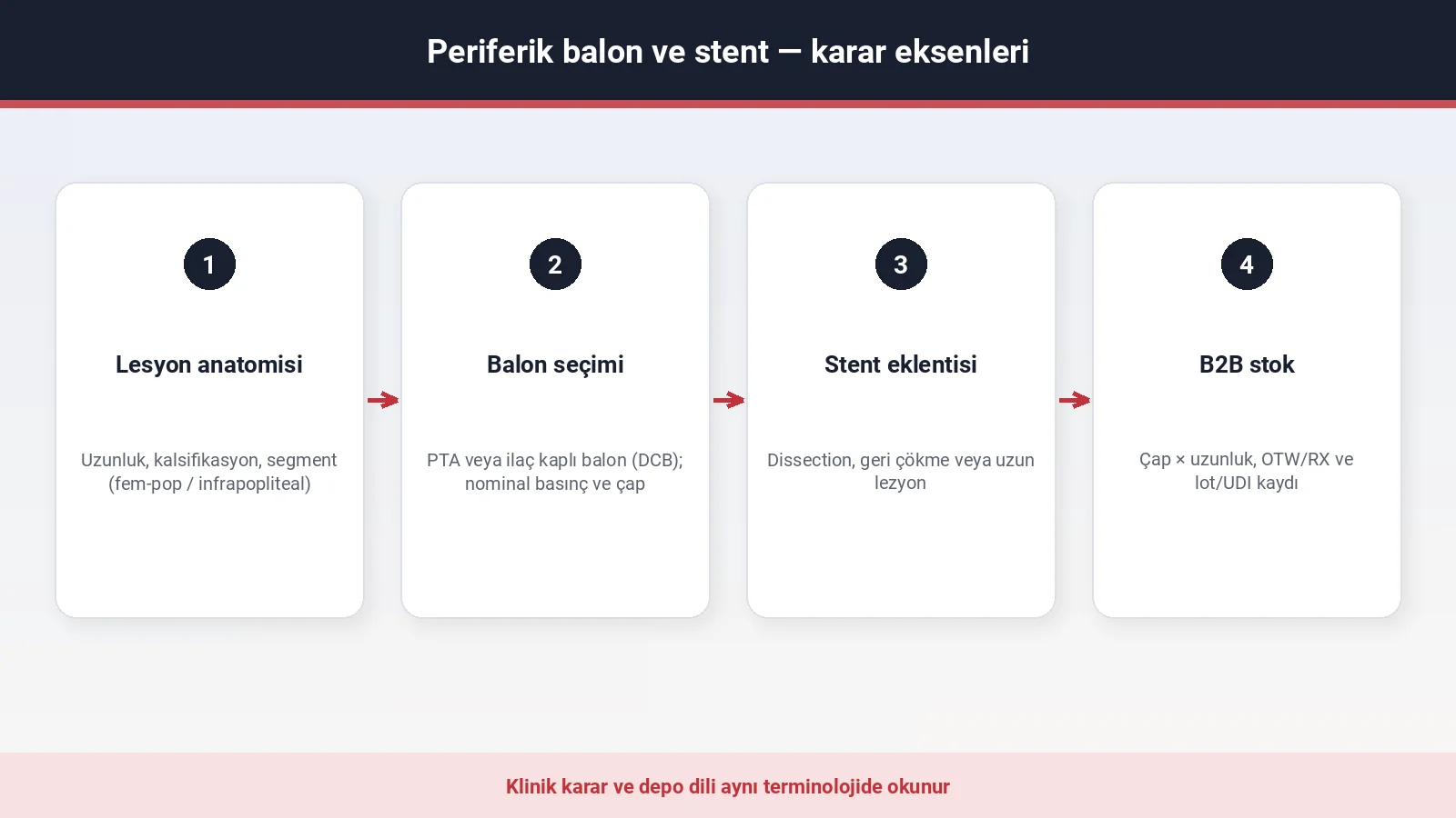
In peripheral arterial disease (PAD), an endovascular procedure may involve balloon angioplasty, a stent, or other vessel preparation options. Lesions do not divide neatly into “a balloon will do” and “a stent is essential”; the segment, the length, calcification, clinical evidence and the behaviour of the vessel wall after the procedure decide the choice. The clinical team uses the same terminology for diameter, length, wire and sheath compatibility and device class.
Lesion type: anatomy shapes the choice
The femoropopliteal and below-the-knee (infrapopliteal) segments have different profiles of movement and calibre. In the femoropopliteal region, flexion and torsion of the popliteal artery can place mechanical stress on a stent. In the infrapopliteal segment the vessel diameter is smaller and lesions may be long and calcified; in a chronic limb-threatening ischaemia (CLTI) profile, the distal target vessel directly affects wound perfusion.
Parameters commonly assessed in clinical practice:
- Lesion length — a short focal stenosis and a long, complex lesion do not fit the same procedural plan.
- Calcification — heavily calcified plaque affects both the nominal pressure needed and the choice of stent.
- Risk of dissection — an intimal tear or recoil after ballooning brings the indication for a stent onto the agenda.
- Distal flow — patency of the target vessel and continuous flow to the foot are assessed together with perfusion of the wound bed in CLTI.
- Previous intervention — restenosis, in-stent restenosis or a stenosis at a bypass anastomosis calls for a different combination of devices.
In a CLTI profile the arterial inflow and the wound are followed in the same file; while wound care continues, infection and perfusion are assessed without delay. That intersection, along with how the medical and endovascular pathways in peripheral arterial disease run alongside one another, is summarised in critical limb ischaemia and wounds.
Balloon options: plain angioplasty and the drug-coated balloon
A percutaneous transluminal angioplasty (PTA) balloon is inflated inside the vessel to widen the lesion. Models without a drug coating are referred to as “plain PTA”; diameter, length and pressure figures are verified from the manufacturer’s instructions for use (IFU). A drug-coated balloon (DCB) is coated with an antiproliferative drug; the decision to use one follows the anatomical segment, the clinical evidence, the approved intended use and the characteristics of the patient. Evidence from the femoropopliteal territory should not be treated as identical to below-the-knee use.
Three measurements are written together when a balloon is specified:
- Diameter (mm) — relative to the reference vessel diameter; undersizing or oversizing affects the technical result.
- Length (mm) — chosen to cover the length of the lesion; a short balloon falls short in a long plaque.
- Nominal pressure (atmospheres) — for calcified lesions, high-pressure models are stated on a separate line in the IFU.
Over-the-wire (OTW) and rapid exchange (RX) systems describe different ways of handling the guidewire; the wire diameter given in the IFU has to be checked against the system. Interventional product groups and abbreviations are summarised in angioplasty product groups.
When the balloon alone is enough
In a short focal stenosis, an acceptable residual stenosis after ballooning, the absence of flow-limiting dissection and adequate distal flow can support leaving the vessel without a stent. Even so, the decision depends on the anatomical territory, the device used and the result of the procedure. Guideline-directed medical therapy continues at every stage; a balloon intervention does not by itself halt the atherosclerotic process. During follow-up, the ankle–brachial index (ABI) and imaging are added to symptoms and vascular examination where needed.
Stenting: when is a stent added?
Dissection, recoil or a long lesion after ballooning may warrant a stent. Peripheral stents are divided into self-expanding and balloon-expandable types; in the femoropopliteal segment and behind the knee, self-expanding nitinol models are commonly used.
How guidelines define the indications for stenting:
- Flow-limiting dissection — an intimal tear after ballooning that restricts flow; a stent or a plan for prolonged inflation.
- Residual stenosis — a significant narrowing remaining after ballooning; most protocols define a threshold value.
- Long lesion or chronic total occlusion (CTO) — the endovascular strategy is planned according to the anatomical territory and the procedural result; length alone does not create a routine obligation to stent.
- In-stent restenosis — recurrent narrowing inside a previous stent; the options of a drug-coated balloon or a further stent layer vary between guidelines.
A coronary stent portfolio should not be confused with peripheral stents; the diameter range, the radial force and the indication in the IFU are different. The product scope is listed on the angioplasty page; the order in which a technical file is read matches the checklist in what to look for in a medical device technical file.
Sizing and compatibility in stent selection
When a stent is ordered, alongside diameter and length the landing zone — the seating of the stent in a healthy segment of vessel — is part of the clinical plan. A short stent falls short in a long lesion; a long stent behind the knee is exposed to flexion stress. The compatible delivery system, sheath size and wire list given in the IFU are checked together with the stent. A stent is not defined without its delivery system.
Guideline recommendations and clinical stage
The 2024 ESC guidelines on peripheral arterial and aortic diseases guide decisions on medical therapy and revascularisation according to the clinical picture and the severity of symptoms. In claudication, medical therapy and structured exercise are the baseline approach; where function remains limited despite this and the anatomy is suitable, revascularisation is considered. In a CLTI profile, revascularisation is considered with the aim of preventing limb loss; wound and infection management run in parallel.
The CLTI framework of the Global Vascular Guidelines — clinical picture, objective measurement of ischaemia, wound and infection burden — positions revascularisation as a means of preventing limb loss. In infrapopliteal revascularisation, the distal target, the lesion anatomy and the available evidence are assessed together. The role of a drug-coated balloon or a stent below the knee cannot be generalised directly from the femoropopliteal territory; a single device type does not suit every anatomy.
The Fontaine or Rutherford classification supports clinical communication; the revascularisation decision is made in the light of anatomy and symptom severity.
Sources
- Mazzolai L, et al. 2024 ESC Guidelines for the management of peripheral arterial and aortic diseases. Eur Heart J. 2024. doi:10.1093/eurheartj/ehae179
- Gerhard-Herman MD, et al. 2016 AHA/ACC Guideline on the Management of Patients With Lower Extremity Peripheral Artery Disease. Circulation. 2017. doi:10.1161/CIR.0000000000000471
- Conte MS, et al. Global Vascular Guidelines on the Management of Chronic Limb-Threatening Ischemia. J Vasc Surg. 2019. doi:10.1016/j.jvs.2019.02.016
- Tepe G, et al. Drug-coated balloon versus standard percutaneous transluminal angioplasty for the treatment of superficial femoral and popliteal peripheral artery disease. Circulation. 2015. doi:10.1161/CIRCULATIONAHA.114.011004
- VascularVita: Angioplasty · Terminology guide · CLTI and wounds · Technical file