Critical limb ischaemia (CLI — chronic limb-threatening ischaemia) often reaches the vascular physician’s desk first, then the wound team’s. An open wound or non-healing ischaemic lesion is the surface expression of inadequate arterial inflow. Changing dressings or moistening the wound bed without a balloon or stent plan may not be enough; if oxygen and nutrients do not reach tissue at the perfusion level, granulation does not progress. This article summarises the wound chain in CLI — from arterial inflow to distal tissue nutrition, from revascularisation decision to wound follow-up — and how two parallel treatment tracks are managed in the same patient.
CLI and the wound chain: from inflow to wound bed
Critical limb ischaemia defines the most advanced stage of peripheral artery disease (PAD), with rest pain, ischaemic wound, or gangrene. In the Global Vascular Guidelines, CLI is defined as “limb-threatening ischaemia”; amputation risk and mortality rise markedly at this stage. The wound is the last link in the chain: flow falls in the proximal vessel, perfusion pressure remains inadequate in the distal segment, tissue oxygenation is impaired, and necrosis or a non-healing wound develops.
In clinical practice, the chain is assessed in this order:
- Risk factors and PAD stage — diabetes, smoking, chronic kidney disease; Fontaine or Rutherford classification.
- Arterial inflow — stenosis or occlusion in femoral, popliteal, and infrapopliteal segments; imaging (duplex, CTA, angiography).
- Distal perfusion — ankle–brachial pressure index (ABPI), toe–brachial index (TBI), transcutaneous oxygen pressure (TcPO2); at wound bed and surrounding skin level.
- Wound bed — ischaemic wound margin, necrotic tissue, infection signs; healing rate.
- Function and offloading — pressure point, foot deformity, walking capacity.
In a venous ulcer profile the problem is often on the outflow side; in CLI the problem is inflow and distal flow. Both mechanisms can coexist in the same leg; the chain cannot then be assessed from one angle only. Perfusion assessment in wound care details the venous versus arterial distinction and the ABPI and TcPO2 measurement.
Revascularisation: the angioplasty perspective
In CLI the goal is to restore adequate limb perfusion and reduce amputation risk. Medical therapy (antiplatelet, statin, smoking cessation) continues at every stage; yet when ischaemic wound or rest pain is present, endovascular or open surgical revascularisation comes into play. The decision is shaped by anatomy, distal target vessel, infection status, and patient comorbidities.
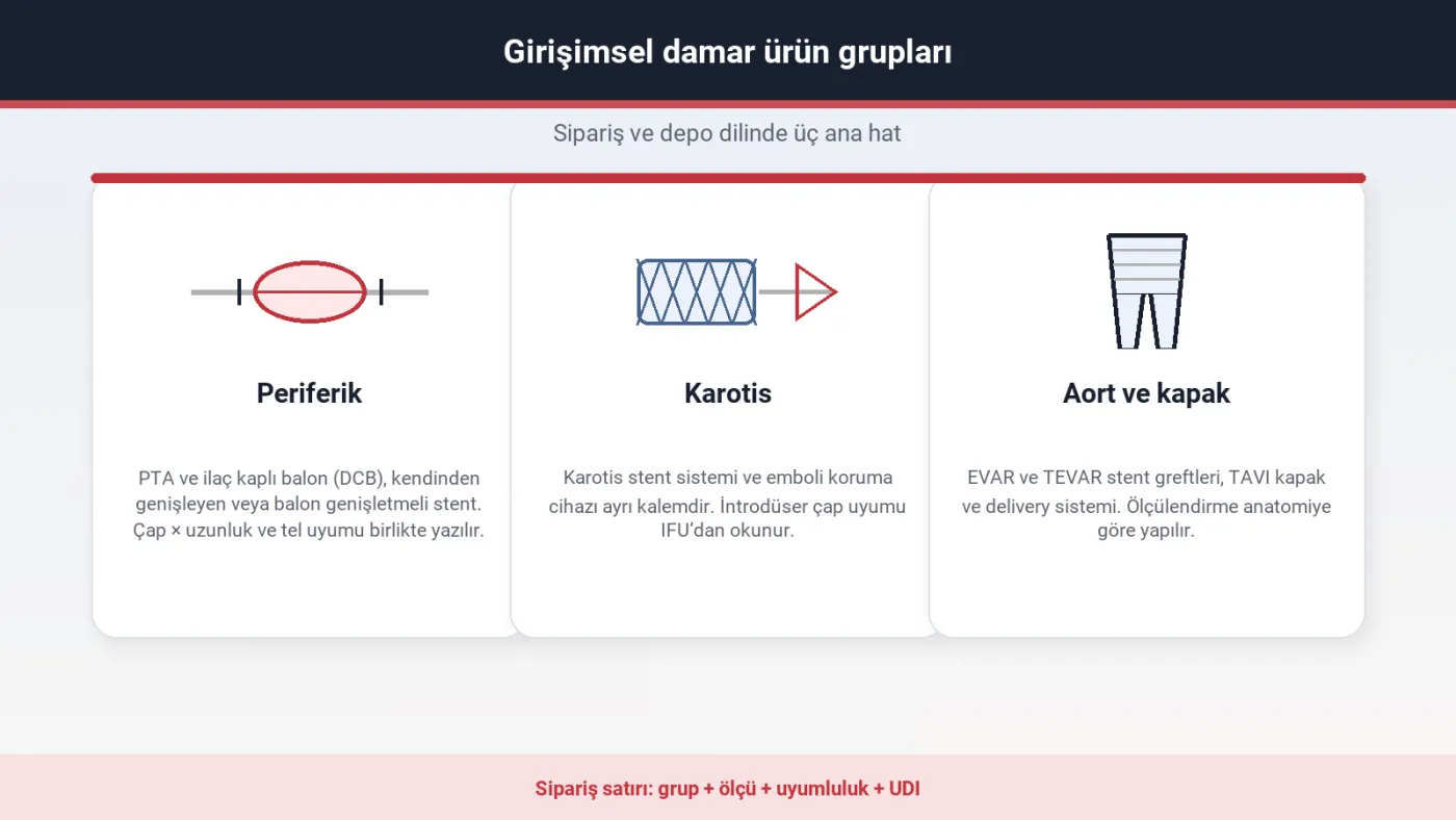
In the endovascular approach, PTA balloon, drug-coated balloon (DCB), and peripheral stent are frequently used line items. In the infrapopliteal segment, vessel diameter may be small and lesions long and calcified; a “single balloon” plan does not fit every scenario. Interventional product terminology — diameter, length, over-the-wire (OTW) or rapid-exchange (RX) systems, and nominal pressure — is terminology shared by the clinical team. Angioplasty product groups summarises peripheral balloon and stent items; product scope is listed on the angioplasty page.
Expectation after revascularisation
Intervention alone does not guarantee wound closure. Granulation may accelerate once distal perfusion improves; yet debridement of necrotic tissue, infection control, and offloading continue in the wound team’s domain. Serial ABPI or TcPO2 measurement in the follow-up protocol provides shared data between the vascular physician and the wound team. The wound may worsen again with restenosis or inadequate technical result; a “vascular work is done” note therefore does not end wound follow-up.
Wound care: once perfusion has been assessed
In the CLI profile the wound is usually distal — toes, pressure points, lateral malleolus. Skin may be cool and pale; the wound margin is sharp, necrotic tissue prominent. Dressings and bandages do not correct arterial inflow; they support the wound bed in an appropriate perfusion environment.
In arterial insufficiency, aggressive moist dressing selection can increase wet necrosis risk in ischaemic tissue. The clinical order of priority is as follows:
- Perfusion and revascularisation plan — if distal oxygenation is inadequate, revascularisation is planned first.
- Infection assessment — colonisation versus infection; antibiotic indication by clinical picture.
- Debridement — necrotic and fibrinous tissue; the plan is reassessed after perfusion improves.
- Offloading — pressure relief; according to foot deformity and gait pattern.
- Dressing category — exudate level and wound bed moisture balance; once the aetiology is established.
Wound care product groups are listed at category level on the wound care page. The aetiology, perfusion, exudate, and infection sequence for dressing selection is summarised in wound dressing assessment. In the CLI patient, “which dressing?” follows “is distal perfusion adequate?”
Two treatment tracks, one follow-up plan
Revascularisation and wound care run as two parallel tracks in the same patient; if notes and meetings remain within separate disciplines, the plan falls apart. Practical workflow converges on these headings:
- Shared patient identity and wound location — a single wound map; photo and measurement date.
- Perfusion data — ABPI/TBI/TcPO2 date and position; before and after revascularisation.
- Intervention record — segment, technique used, technical file/lot; restenosis follow-up.
- Wound progress — surface area, granulation quality, infection score.
- Function — pain, walking distance, offloading adherence.
Vascular health relates in systemic context to the healthspan discussion: peripheral artery disease is part of the cardiovascular risk profile. Vascular health and healthspan discusses vascular integrity in this context; programme-level context is on the longevity page.
Common pitfalls
Focusing only on the wound surface can hide arterial inflow. Even when pulse palpation feels “normal,” distal perfusion may remain inadequate in the diabetic foot. Ending wound follow-up early after revascularisation misses restenosis. When venous oedema and arterial ischaemia coexist, the compression plan must align with ABPI; detail is summarised in compression pressure (mmHg).
Sources
- Conte MS, et al. Global Vascular Guidelines on the Management of Chronic Limb-Threatening Ischemia. J Vasc Surg. 2019. doi:10.1016/j.jvs.2019.02.016
- Aboyans V, et al. ESC Guidelines on peripheral arterial diseases. Eur Heart J. 2024. doi:10.1093/eurheartj/ehae179
- International Working Group on the Diabetic Foot (IWGDF). Guidelines on peripheral artery disease. 2023. iwgdfguidelines.org
- Snyder RJ, et al. Wound Healing Society update on guidelines for arterial ulcers. Wound Repair Regen. 2016. doi:10.1111/wrr.12348
- VascularVita: Angioplasty · Wound care · Perfusion assessment · Longevity