
Diabetic foot wounds are often seen in primary care or the emergency department; before the patient reaches a wound team or vascular surgeon, the question “which dressing?” comes up. Yet diabetic foot disease lies at the intersection of neuropathy, arterial ischaemia, infection, and pressure load; dressing choice is the last step in that picture. In VascularVita wound care, aetiology and perfusion are assessed first; referral decisions rest on that data. This article summarises the sequence of first assessment in diabetic foot wounds, when to consider referral to which specialist, and where dressing category enters the conversation.
First assessment: four domains
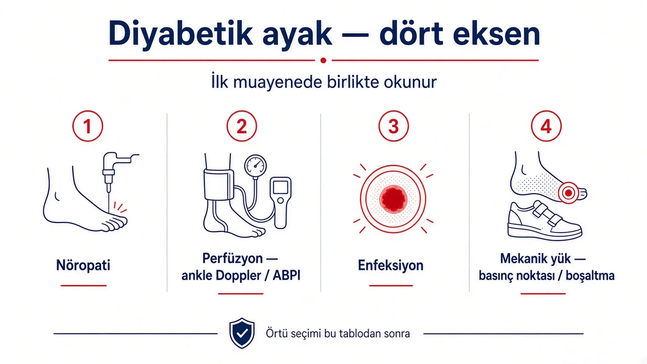
In diabetic foot examination, four domains are assessed together. A single finding is not reduced to “simple wound” or “infection.”
- Neuropathy — monofilament test, vibration sense, foot deformity; loss of pain cue prolongs wound duration.
- Arterial perfusion — pulse palpation, ankle–brachial pressure index (ABPI); in diabetes, medial calcification can mislead the index.
- Infection — erythema, warmth, purulent exudate, osteomyelitis signs; colonisation is separated from infection.
- Mechanical load — pressure point, footwear fit, offloading need; Charcot foot deformity is assessed separately.
Wound size, depth, and exudate level are added on top of these four domains. A wound map — site, duration, prior treatment — is kept in one note; the same reference is used in follow-up visits. The questions to ask before dressing selection are summarised in before choosing a wound product.
Perfusion and ischaemia: the inflow side first
Arterial ischaemia is common in diabetic foot; distal perfusion may remain inadequate even when the pulse feels “normal.” ABPI is useful for screening; values below 0.9 or above 1.3 require further testing. In a suspicious profile, toe–brachial index (TBI) or transcutaneous oxygen pressure (TcPO2) provides complementary information.
In an ischaemic wound profile, skin may be cool and pale; the wound margin is sharp, necrotic tissue prominent. Here the priority is revascularisation or a vessel-sparing plan; moist dressings in ischaemic tissue can increase wet necrosis risk. Focusing on the wound surface before perfusion has been assessed does not allow realistic expectations of healing. Venous oedema and arterial ischaemia can coexist in the same leg; both components are assessed separately. Further detail is given in perfusion assessment in wound care.
When ABPI can be misleading
- Chronic kidney disease and long-standing diabetes — medial calcification.
- Distal wound (toes) — ankle pressure may not reflect toe perfusion.
- Severe oedema — cuff placement is difficult; measurement is repeated.
When the picture intersects critical limb ischaemia (CLI), vascular intervention and wound follow-up run in parallel. Critical limb ischaemia and wounds summarises joint follow-up for both treatment tracks.
Referral: which specialist, which finding
After first assessment, referral is shaped by findings. A single “diabetic foot clinic” may not cover every scenario; profiles requiring urgent referral are recognised separately.
- Vascular surgery / interventional physician — low ABPI or TBI, rest pain, ischaemic wound, gangrene; revascularisation assessment.
- Orthopaedics / foot surgery — deep infection, suspected osteomyelitis, Charcot deformity, surgical debridement or offloading device need.
- Wound care team — chronic wound follow-up, debridement plan, exudate management; after perfusion and infection are clear.
- Endocrinology / internal medicine — glycaemic control, foot examination frequency; long-term risk reduction.
Findings requiring urgent referral: rapidly progressing infection, systemic inflammatory response, ischaemic gangrene, suspected sepsis. In this profile, dressing change is not deferred; yet the urgent field remains limited to infection and perfusion — a product catalogue does not enter the room.
A referral note summarises wound photograph date, ABPI/TBI value and position, infection findings, and offloading status on one page. The next specialist does not have to re-collect the same data; patient confidence and time loss decrease.
Infection versus colonisation
Colonisation is common on the diabetic foot; not every profile warrants an antimicrobial dressing or systemic therapy. Infection is assessed against the clinical picture — periwound erythema, warmth, purulent exudate, progressive wound enlargement. When osteomyelitis is suspected, imaging and orthopaedic consultation enter the plan.
Antimicrobial dressing category is considered once infection has been confirmed, in line with clinical guideline and institutional protocol. Routine antimicrobial dressing for colonisation alone is not required; IWGDF and wound community guidelines emphasise this distinction. Dressings do not replace infection treatment; surgical and antibiotic plans run separately.
Dressing category: once the aetiology is established
Dressings and bandages in diabetic foot are selected once the four domains have been assessed. Category-level choice rests on these questions:
- Exudate level — dry, low, moderate, or high; moisture balance is chosen from this data.
- Wound bed — necrotic tissue, fibrin, granulation; debridement need is planned separately.
- Periwound skin — maceration, xerosis; periwound protection product groups may enter the plan.
- Offloading — pressure relief may be independent of dressing and take priority.
Product groups are listed at category level on the wound care page; When a venous oedema component is present, compression is assessed after adequate arterial inflow is confirmed; pressure grades and ABPI summary are in compression pressure (mmHg).
Dressing approach in ischaemic profile
When distal perfusion is inadequate, the wound surface may be kept dry; aggressive moist dressings increase risk in ischaemic tissue. After revascularisation the wound bed is reassessed; when granulation accelerates, moisture balance is updated. This order reverses the “dressing first, vessels later” flow.
Dressing approach in neuropathic profile
When neuropathy is present, pain cues are lost and wound duration lengthens. Offloading and regular foot examination come before dressing choice. Exudate level and infection signs are assessed separately in neuropathic profiles as well; “the patient feels no pain” is not used to skip perfusion and infection screening.
Clinical flow summary at first examination
Practical sequence is summarised as follows:
- Four-domain screen — neuropathy, perfusion, infection, mechanical load.
- ABPI or TBI; TcPO2 in suspicious profiles.
- Referral decision — vascular, orthopaedic, wound team, emergency.
- Offloading and glycaemic control plan.
- Dressing category — exudate, wound bed, periwound skin.
- Follow-up date and shared wound map update.
Vascular health in diabetic foot wounds is summarised in systemic context in vascular health and healthspan; peripheral artery disease and the wound chain are followed in the same patient file.
Sources
- International Working Group on the Diabetic Foot (IWGDF). Guidelines on the prevention and management of diabetic foot disease. 2023. iwgdfguidelines.org
- Schaper NC, et al. IWGDF Guideline on the diagnosis and management of foot infections in persons with diabetes. Diabetes Metab Res Rev. 2023. doi:10.1002/dmrr.3651
- Conte MS, et al. Global Vascular Guidelines on the Management of Chronic Limb-Threatening Ischemia. J Vasc Surg. 2019. doi:10.1016/j.jvs.2019.02.016
- Hinchliffe RJ, et al. IWGDF Guideline on the diagnosis and management of peripheral artery disease in patients with diabetes. Diabetes Metab Res Rev. 2023. doi:10.1002/dmrr.3650
- VascularVita: Wound care · Before choosing a wound product · Compression pressure