In the VascularVita longevity programme, vascular health is monitored as one of the pillars of function. How far a person can walk, how fast a wound heals and organ reserve depend largely on perfusion — blood flow reaching the tissue. Behind perfusion sit the arterial wall, the small-vessel network (microcirculation) and the endothelium that lines the vessel. Low-grade chronic inflammation that rises with age (inflammaging) strains all of these layers at once. The result is not a single laboratory value; it is seen in walking capacity, wound healing and daily function.
This article summarises where inflammation meets vascular ageing, from the patient’s clinical picture and treatment. Disrupted signalling between cells is described in altered intercellular communication, and mitochondrial stress in mitochondrial dysfunction. The focus here is the link between endothelium, vessel wall and perfusion.
Inflammaging: background inflammation, not infection
Inflammaging is low-grade, persistent inflammation that rises with age without an infection. Markers such as interleukin-6 (IL-6) and C-reactive protein (CRP) stay mildly but persistently raised. The sources are several: visceral fat, a weakened gut barrier, oxidative stress, and inflammatory molecules secreted by senescent cells (SASP: senescence-associated secretory phenotype). The load does not stay in one organ; it affects the whole vascular bed.
A single raised CRP does not establish a diagnosis in clinical practice. The course of the value over time is interpreted alongside the clinical picture. In longevity follow-up the aim is not to pull one marker into the reference range, but to see how far inflammation limits perfusion and function.
The endothelium: the vessel’s regulatory surface
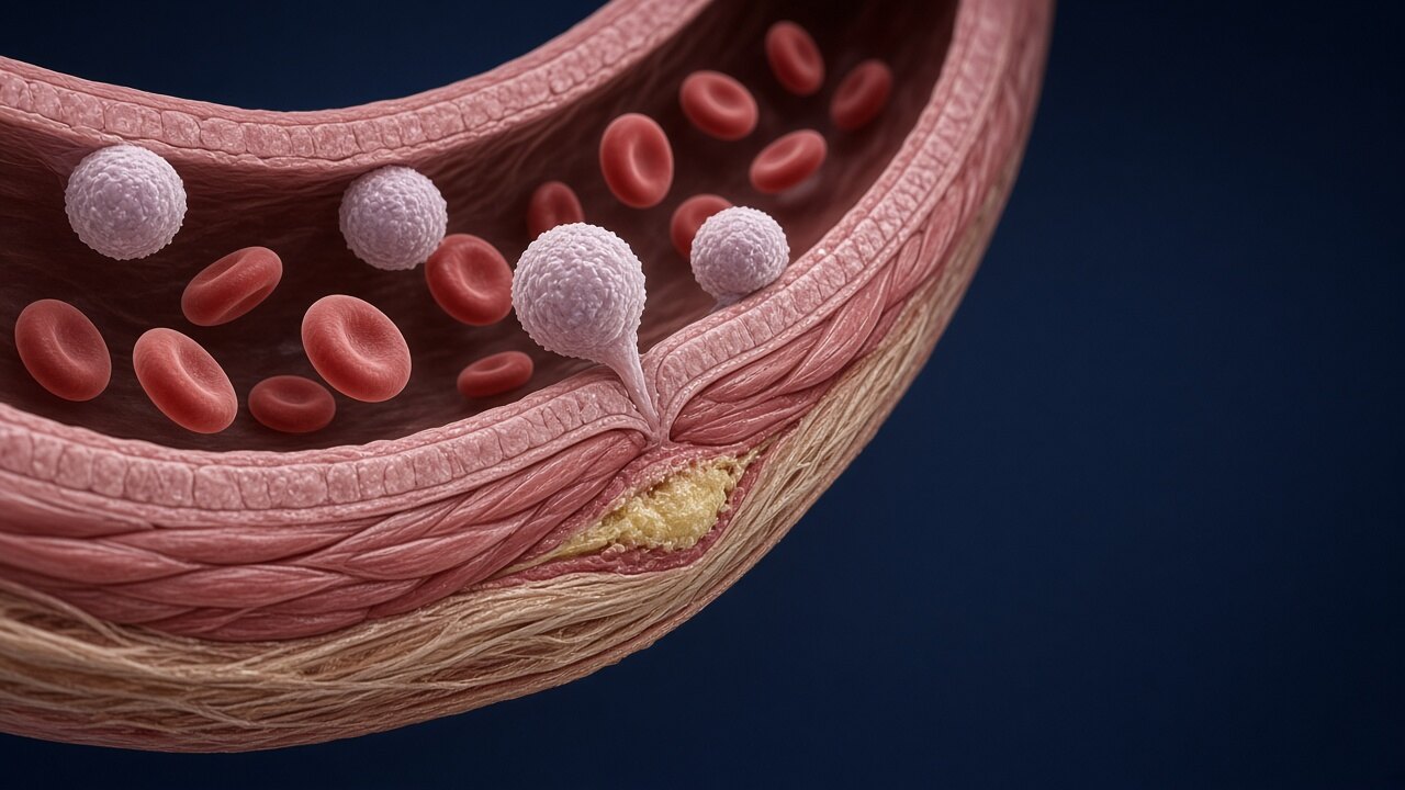
The endothelium is the single cell layer between blood and vessel wall. Through the nitric oxide (NO) it produces, it supports vasodilation and limits platelet adhesion and the passage of white cells into the wall. When inflammation rises, adhesion molecules (E-selectin, VCAM-1, ICAM-1) increase on the endothelial surface and NO production falls. Monocytes adhere to that surface and enter the wall. That is the first step of atherosclerosis. Capillaries and small arterioles are affected by the same process.
Vascular ageing is measured in different ways, and each method shows a different layer. Pulse wave velocity (PWV) reflects arterial stiffness, carotid intima–media thickness (IMT) wall thickening, and flow-mediated dilation (FMD) endothelial function. These measurements do not replace one another; they are interpreted together.
Vascular ageing and perfusion
Vascular ageing covers endothelial dysfunction, wall thickening, loss of elastic fibres and a fall in capillary number (microvascular rarefaction). When perfusion falls, muscle repair is delayed, the wound bed is not oxygenated enough, and kidney and brain reserve narrow. In chronic limb-threatening ischaemia (CLTI), arterial flow and the wound become the same clinical problem. That chain is described in critical limb ischaemia and wounds.
Targeting inflammation: what does the evidence say?
The role of inflammation in vascular disease does not rest on observational data alone. In the CANTOS trial, canakinumab targeting IL-1β reduced recurrent cardiovascular events without lowering cholesterol, in patients with a prior myocardial infarction and a raised CRP. In LoDoCo2, low-dose colchicine lowered cardiovascular event risk in people with chronic coronary disease. Both trials place inflammation among the causes of atherosclerotic events. Those results were obtained in diagnosed vascular disease; they are not evidence for preventive anti-inflammatory medicines in a healthy older adult.
Clinical findings: walking, wounds, function
Inflammation and vascular ageing show themselves in concrete findings:
- Intermittent claudication: Calf pain on walking and a shorter walking distance. An early finding of peripheral artery disease.
- Delayed wound healing: Inadequate perfusion and persistent inflammation are assessed together.
- Decline in intrinsic capacity: A mild fall at the same time in locomotion, cognition and vitality in the WHO ICOPE framework.
- Frailty: The picture where inflammation, loss of muscle and a fall in vascular reserve meet. Early findings are described in frailty.
These findings are not, on their own, a diagnosis of “vascular ageing”; they are the vascular-layer reflection of lost function. When needed, vascular surgery, cardiology and the wound team work in the same record.
Management: prevention, treatment, follow-up
Prevention. The steps with the strongest evidence are regular physical activity, stopping smoking, weight and glycaemic control, and enough sleep. These steps lower inflammation markers and improve endothelial function.
Indicated treatment. In diagnosed vascular disease, antiplatelet and lipid-lowering treatment continues according to the guideline. In peripheral artery disease the revascularisation decision follows anatomy and symptom severity. The order of medical and endovascular treatment is described in the medical and endovascular pathway in peripheral artery disease. The wound team does not move to a dressing until perfusion is clear.
Follow-up. Walking distance, the wound and daily function are reassessed at set intervals. Inflammation markers support that follow-up; they do not replace it. The flow is collected on the individual assessment form.
Summary
Inflammaging is a persistent inflammatory load that impairs the endothelium’s regulatory function and the microcirculation. Randomised trials support a causal role for inflammation in vascular disease. Loss of perfusion presents in clinical practice as a shorter walking distance, delayed wounds and a decline in function. Management is lifestyle measures, guideline-based vascular treatment and regular functional follow-up.
Sources
- López-Otín C, Blasco MA, Partridge L, Serrano M, Kroemer G. Hallmarks of aging: an expanding universe. Cell. 2023;186(2):243–278. doi:10.1016/j.cell.2022.11.001
- Franceschi C, Garagnani P, Parini P, Giuliani C, Santoro A. Inflammaging: a new immune–metabolic viewpoint for age-related diseases. Nat Rev Endocrinol. 2018;14(10):576–590. doi:10.1038/s41574-018-0059-4
- Ferrucci L, Fabbri E. Inflammageing: chronic inflammation in ageing, cardiovascular disease, and frailty. Nat Rev Cardiol. 2018;15(9):505–522. doi:10.1038/s41569-018-0064-2
- Libby P, Ridker PM, Hansson GK. Inflammation in atherosclerosis: from pathophysiology to practice. J Am Coll Cardiol. 2009;54(23):2129–2138. doi:10.1016/j.jacc.2009.09.009
- Ridker PM, et al. Antiinflammatory therapy with canakinumab for atherosclerotic disease (CANTOS). N Engl J Med. 2017;377:1119–1131. doi:10.1056/NEJMoa1707914
- Nidorf SM, et al. Colchicine in patients with chronic coronary disease (LoDoCo2). N Engl J Med. 2020;383:1838–1847. doi:10.1056/NEJMoa2021372
- Cesari M, et al. Evidence for the domains supporting the construct of intrinsic capacity. J Gerontol A Biol Sci Med Sci. 2018;73(12):1653–1660. doi:10.1093/gerona/gly011