In VascularVita longevity work, the aim is not to reverse chronological age. The aim is to protect the years in which a person can walk, decide, and sustain daily life — healthspan. Ageing is not reduced to a single test. Cellular and molecular decline, organ-system change, a person’s intrinsic capacity and daily function form a single continuum. Management follows from this assessment: define the clinical picture, reduce risk, give indicated treatment and monitor.
At the cellular level, one of the integrative drivers in this continuum is stem cell exhaustion. In López-Otín and colleagues’ framework of the hallmarks of aging, this hallmark covers the quantitative and qualitative loss of tissue regenerative reserve. Senescent cells, epigenetic drift, and intercellular communication disrupt the niche; this article explains the regenerative pool. All nine hallmarks appear in the nine hallmarks of ageing; the programme is described in the ageing stages and in individual assessment.
What are stem cells and the niche?
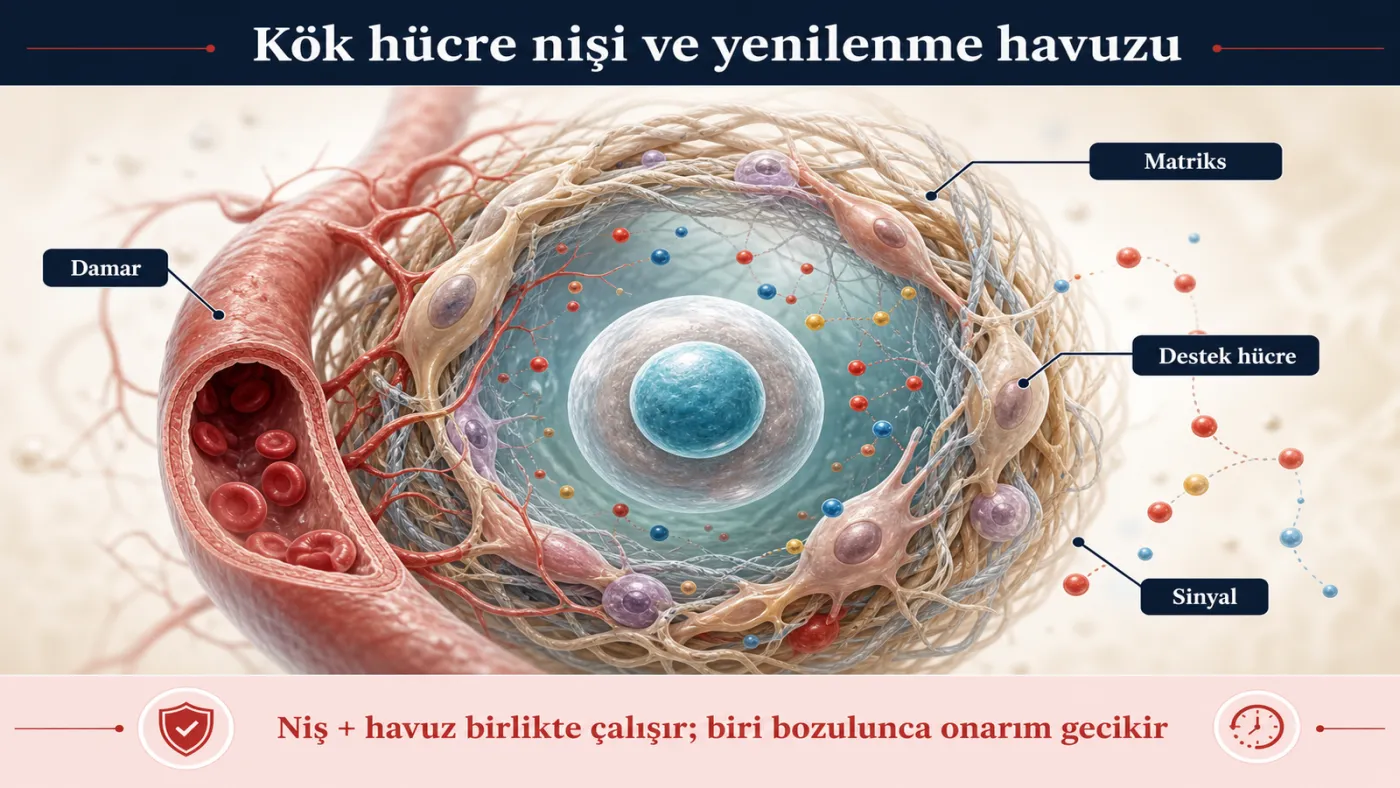
Adult tissues rely on stem cells and progenitor cells to repair damage. These cells self-renew and, when needed, differentiate into specialised cells. That alone is not enough: a stem cell lives inside a niche — neighbouring support cells, the vascular bed, matrix, and secretory signals. When the niche is intact, the pool stays balanced; when it is disrupted, proliferation, differentiation, or the quiescent state is set incorrectly.
Examples differ by tissue. Haematopoietic stem cells (HSCs) produce blood and immune cells. Muscle satellite cells contribute to repair after injury. Continuous renewal is also required in skin and intestinal epithelium. In a longevity assessment, the issue is not “increasing stem-cell numbers”; it is niche and pool working together (López-Otín 2023).
Why does the balance break with age?
With age, the stem-cell pool narrows: numbers may fall, function may decline, or differentiation may drift in the wrong direction. The niche also deteriorates. Accumulation of senescent cells and the SASP suppress repair through inflammatory signals. Genomic damage, telomere stress, and epigenetic drift strain the stem cells’ own genome and expression programme. Mitochondrial and proteostasis load also consume the same reserve.
The result is not a single laboratory measure. Tissue cannot close damage at the same pace; immune response weakens or shifts; muscle and wound repair slow. In this integrative hallmark, the effects of the other hallmarks become clinically visible as reduced regenerative capacity.
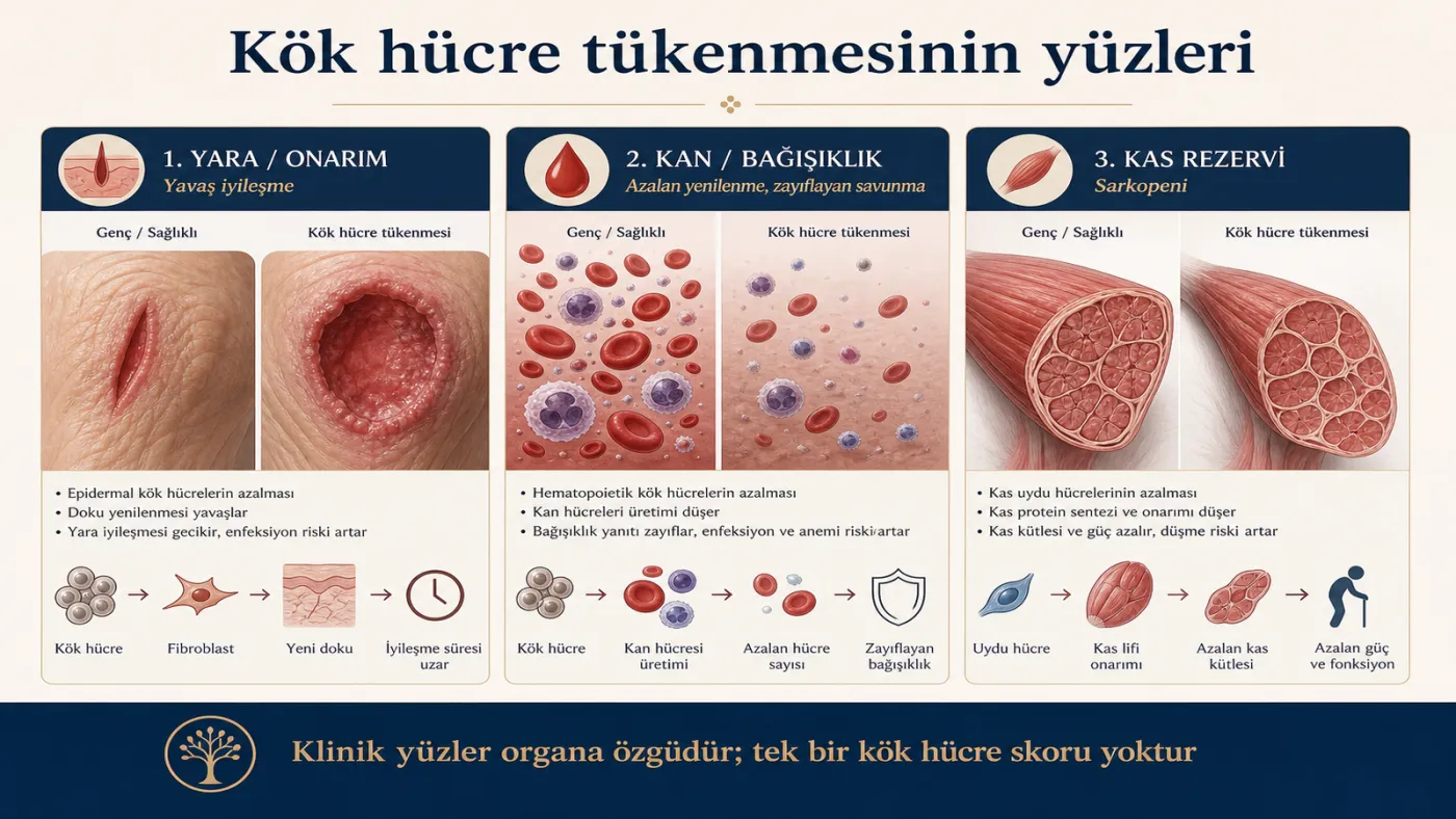
Clinical face: wound, blood, muscle, immunity
Clinical manifestations include delayed wound healing, haematopoietic changes, sarcopenia, and immunosenescence (immune ageing). Chronic wounds and critical limb ischaemia functionally resemble a “regenerative exhaustion” phenotype: when perfusion and niche fail, repair does not complete. These belong to organ-specific disease management; they are not a “stem-cell score” diagnosis.
The World Health Organization defines healthy ageing not as the absence of disease but as the preservation of functional ability; the same principle is developed in our article on healthspan. In longevity management, walking, grip strength, wound-closure rate, and daily independence are functional proxies.
What can be done?
The most mature lever is to protect the niche. Resistance exercise, adequate protein, sleep, and metabolic–inflammatory control support the base for muscle and systemic renewal. Sound nutrition supports the same network; this overlaps with nutrient sensing and proteostasis.
In disease indications, mesenchymal stem cell (MSC) and similar regenerative approaches are clinical options governed by guidelines and regulation; they are not a general longevity offering. In research, partial epigenetic reprogramming, plasma / youthful factors, and extracellular vesicle (EV / exosome)–based signals are studied. For technical details, see the exosome technical file article. Senolytic approaches aim to ease the niche indirectly; the evidence level remains research-stage.
What is done in management?
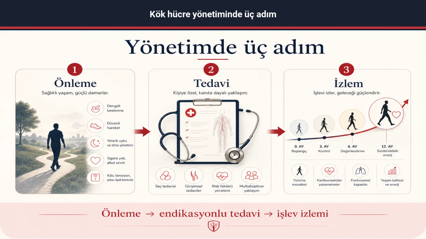
In the VascularVita approach, stem cell exhaustion is addressed in three concrete steps.
First is prevention. Resistance exercise, adequate protein, sound nutrition, sleep, and inflammatory–metabolic risk control support the niche. The aim is not to “measure renewal with a score”; it is to keep reserve and repair capacity upright.
Second is disease-guided treatment. Chronic wound, vascular involvement, unexplained blood-count disturbance, or sarcopenia is referred to the relevant specialty as the clinical picture requires. Indicated treatment follows the guideline. Longevity screening assesses the ICOPE domains, frailty, and daily function together; it does not assign a stage from a single “stem-cell score.”
The third step is monitoring over time. Loss of renewal is linked to senescent-cell accumulation and communication failure. That is why attention is not limited to a single laboratory result. The same person’s walking, grip strength, wound-healing rate, and independence are reassessed at defined intervals. The flow is on the individual assessment and ageing stages pages.
Summary
Stem cell exhaustion is an integrative hallmark of ageing that arises when the regenerative pool and the niche deteriorate together. Senescent-cell secretion, epigenetic drift, and inflammation narrow the same reserve. The most mature intervention is exercise, protein–sleep, and niche protection. MSC and plasma / EV approaches are indication- or research-bound. In VascularVita longevity management, this hallmark is addressed through prevention, indicated treatment, and function-focused monitoring.
References
- López-Otín C, Blasco MA, Partridge L, Serrano M, Kroemer G. The hallmarks of aging. Cell. 2013;153(6):1194–1217. doi:10.1016/j.cell.2013.05.039 · PMC3836174
- López-Otín C, Blasco MA, Partridge L, Serrano M, Kroemer G. Hallmarks of aging: an expanding universe. Cell. 2023;186(2):243–278. doi:10.1016/j.cell.2022.11.001
- Coppé JP, Desprez PY, Krtolica A, Campisi J. The senescence-associated secretory phenotype: the dark side of tumor suppression. Annu Rev Pathol. 2010;5:99–118. doi:10.1146/annurev-pathol-121808-102144 · PMC2885946
- Kennedy BK, Berger SL, Brunet A, et al. Geroscience: linking aging to chronic disease. Cell. 2014;159(4):709–713. doi:10.1016/j.cell.2014.10.039 · PMC4852871
- World Health Organization. World report on ageing and health. Geneva: WHO; 2015. who.int
- Cesari M, Araujo de Carvalho I, Amuthavalli Thiyagarajan J, et al. Evidence for the domains of the WHO’s Integrated Care for Older People (ICOPE). J Gerontol A Biol Sci Med Sci. 2018;73(12):1653–1660. doi:10.1093/gerona/gly011
- Fried LP, Tangen CM, Walston J, et al. Frailty in older adults: evidence for a phenotype. J Gerontol A Biol Sci Med Sci. 2001;56(3):M146–M156. doi:10.1093/gerona/56.3.m146
Related articles
- Senescent cells (cellular senescence)
- Altered intercellular communication
- Epigenetic alterations: gene expression and ageing
- Mitochondrial dysfunction: energy and ageing
- Exosome technical file
- The nine hallmarks of ageing
- Healthspan
- ICOPE and intrinsic capacity
- Frailty and early findings
- Healthy longevity · Ageing stages · Individual assessment